The accessible volume#
Fluorophores are usually attached to a biomolecule of interest via a relatively flexible, aliphatic spacer arm. This carbon linker endows the fluorophore with motional freedom which is the basis for the common assumption of near isotropic dye rotation, expressed by an orientation factor \(\kappa=2/3\). At the same time, the flexibility of the dye linker introduces some uncertainty in pinpointing the average position of the dye with respect to the biomolecule.
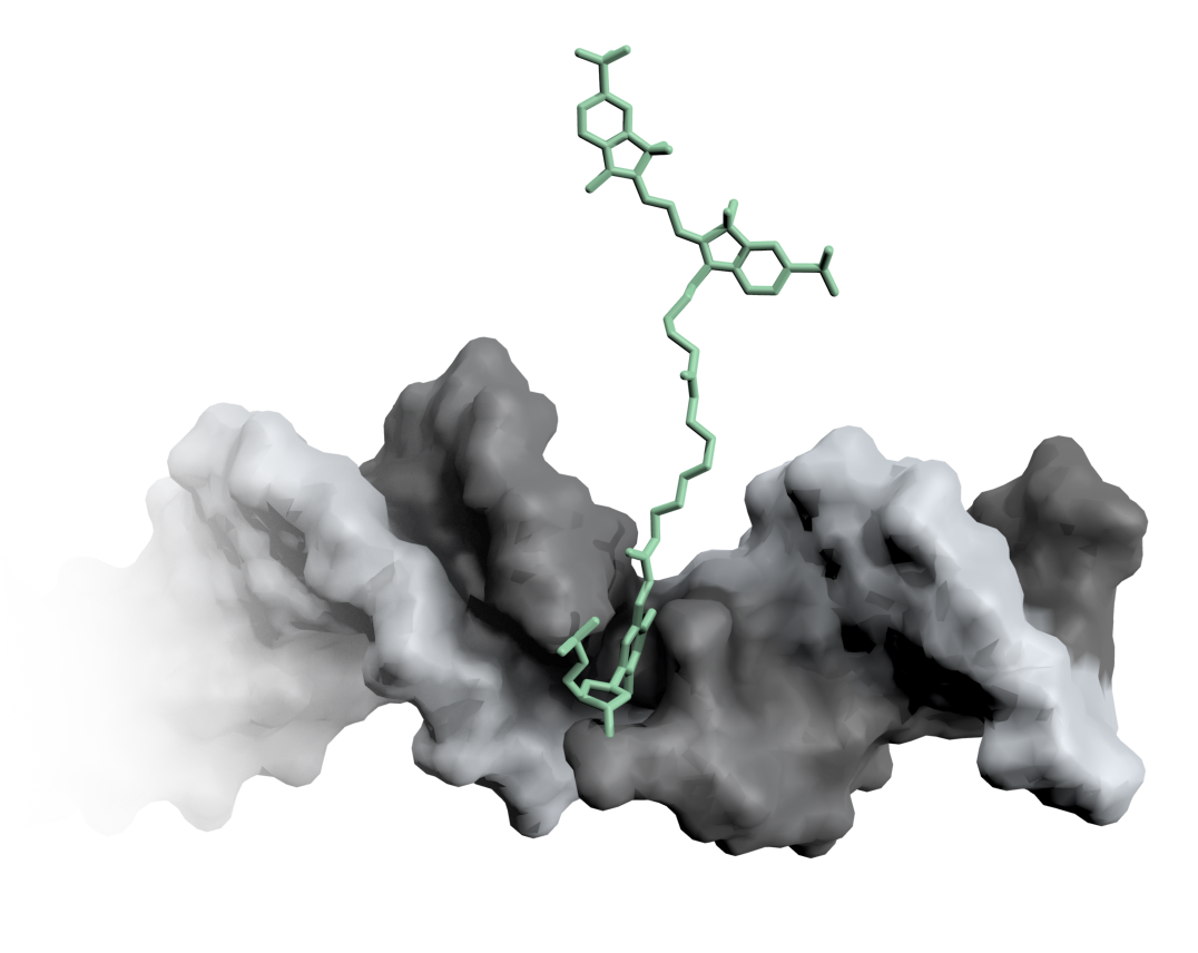
Fig. 4 A Cy3 fluorophore is coupled covalently to a DNA double helix.#
Molecular dynamics simulations with explicitly included fluorophores can sample the dye distribution around a biomolecule. [6, 7, 8] Yet, these simulations are computationally fairly expensive. The accessible volume (AV) approach mitigates this problem by estimating the dye distribution with a purely geometrical search algorithm.
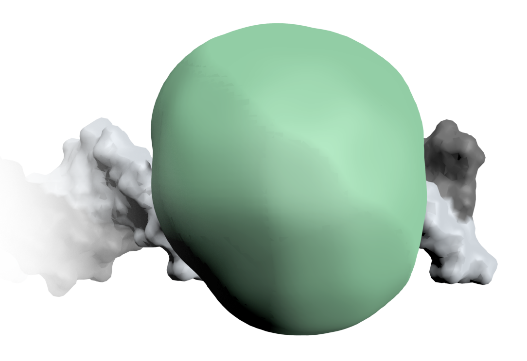
Fig. 5 The dye mobility on the DNA is estimated by the accessible volume.#
In the accessible volume framework the dye is approximated by a spheroid (defined by three radii \(R_1-R_3\)). This dye probe is set to explore the space around the biomolecule within a radius defined by the linker length without clashing with the surface of the biomolecule. [4, 9]
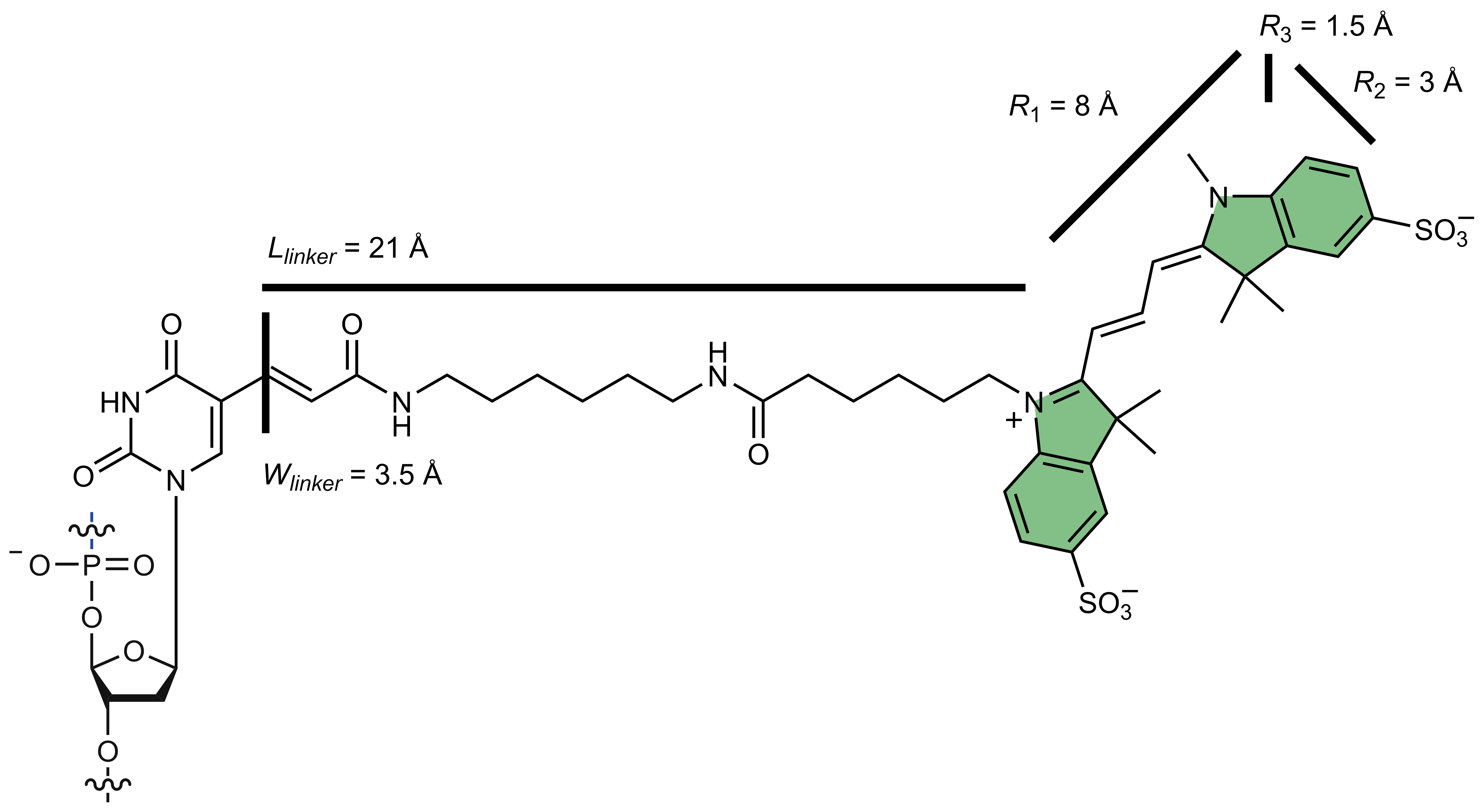
Fig. 6 The Cy3 dye is attached to a deoxyuracil via a flexible carbon linker. The dye and the spacer are parameterized by five distances (\(R_1-R_3\), \(L_\text{linker}\) and \(W_\text{linker}\))#
The maximal distance from the attachment position given by the linker dimensions. The possible dye positions are updated iteratively using a Dijkstra shortest path algorithm. The resulting point cloud describes the location of the dye relative to a static biomolecule.